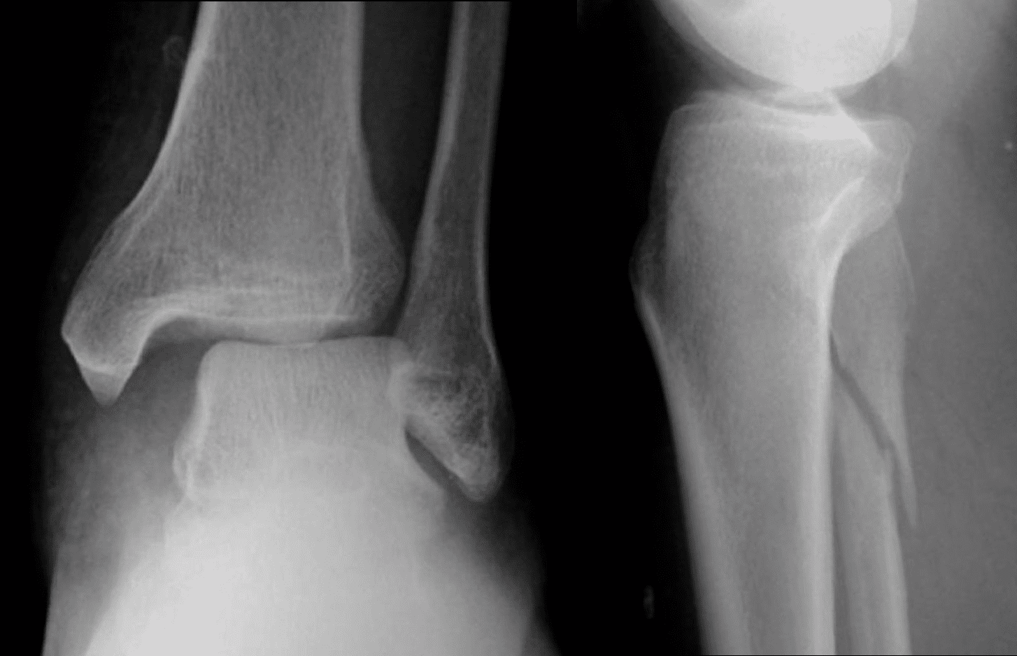
Stress fractures occur in bones that undergo mechanical fatigue. These are caused by repeated trauma from prolonged walking. Stress fractures of the metatarsals are known as a 'march' fracture. These fractures do not require reduction or immobilisation. The phalanges and metatarsals are commonly injured by a heavy object falling onto the foot. Such fractures can be very painful and may be accompanied by vertebral fractures. Fractures of the calcaneum often occur as a result of a fall from height. The bony anatomy of the foot means that a variety of fractures are possible. If this cannot be achieved by manipulation and plaster cast, an individual may have to undergo an open reduction with internal fixation (ORIF) followed by immobilisation in a plaster cast. If displaced, a surgeon should reduce the fragments to the normal anatomical position of the joint. Disruption to articular surfaces may lead to the development of osteoarthritis.įractures without displacement can be treated with a below knee cast that may be applied for 3-6 weeks. Examples include the Danis-Weber classification which identifies the level of injury and the Lauge-Hansen classification which is based on the mechanism of the injury which have predictable patterns and imaging findings.Ĭomplications of ankle fractures can include reduced range of motion at the ankle and foot joints, as a result of peri-articular and intra-articular adhesions or disruption of articular surfaces. Several classifications exist and are used to determine the severity of injury and management. Ottawa rules provide clinicians with a tool to decide whether the joint should be imaged or not. Ankle fractures are most commonly diagnosed through clinical examination and x-ray. The lateral tib/fib demonstrates the proximal fibula fracture and several bony fragments anterior to the ankle joint. There is an oblique/spiral fracture of the proximal fibula. This injury is suggestive of a proximal fibula fracture (Maisonneuve fracture). The mortise view demonstrates a transverse fracture of the medial malleolus and widening of the tibiofibular syndesmosis. On examination he had a painful ankle and was unable to weightbear. This 24 year old male presented to the ED after rolling his ankle. inferior transverse tibiofibular ligament (TrTFL).posterior- inferior tibiofibular ligament (PiTFL).anterior-inferior tibiofibular ligament (AITFL).syndesmosis is made up of 4 ligaments which stabilize the distal tibiofibular syndesmosis: However, when eversion injury occurs, there is often substantial damage to bony and ligamentous supporting structures and loss of joint stability.Ĭhronic Tibiofibular Syndesmosis Injury of Ankle: Evaluation with Contrast-enhanced Fat-suppressed 3D Fast Spoiled Gradient-recalled Acquisition in the Steady State MR Imaging As a result, the ankle is more stable and resistant to eversion injury than inversion injury. Second, the deltoid ligament stabilizing the medial aspect of the ankle joint offers stronger support than the thinner lateral ligaments.


First, the medial malleolus is shorter than the lateral malleolus, allowing the talus to invert more than evert.Inversion and eversion occur at the subtalar joint.Įxcessive inversion stress is the most common cause of ankle injuries for 2 anatomic reasons. The primary motion of the ankle at the true ankle joint (tibiotalar joint) is plantarflexion and dorsiflexion. This is because of the presumed disruption of all the syndesmotic ligaments and the interosseous membrane from the ankle joint to the level of the fibula fracture. The Maisonneuve fracture of the fibula is often considered one of the most unstable ankle injuries. The Maisonneuve fracture consists of a proximal fibular fracture with associated ligament disruption or medial malleolar ankle fracture. When a syndesmosis injury is demonstrated on ankle radiography, the radiographer should consider the possibility of a Maisonneuve fracture and undertake supplementary views to demonstrate the entire lower leg. Radiographers should be aware of the Maisonneuve fracture, its mechanism of injury and the potential for an asymptomatic proximal fibular fracture. The proximal fibular fracture is often not appreciated because of the distracting ankle injury. Maisonneuve fractures are commonly missed clinically and radiographically.


 0 kommentar(er)
